
Dual-Plane Breast Augmentation: Placement, Benefits and Tradeoffs
See where a dual-plane implant sits, how Types I–III differ, the limits for sagging, and questions to ask about animation and implant position.
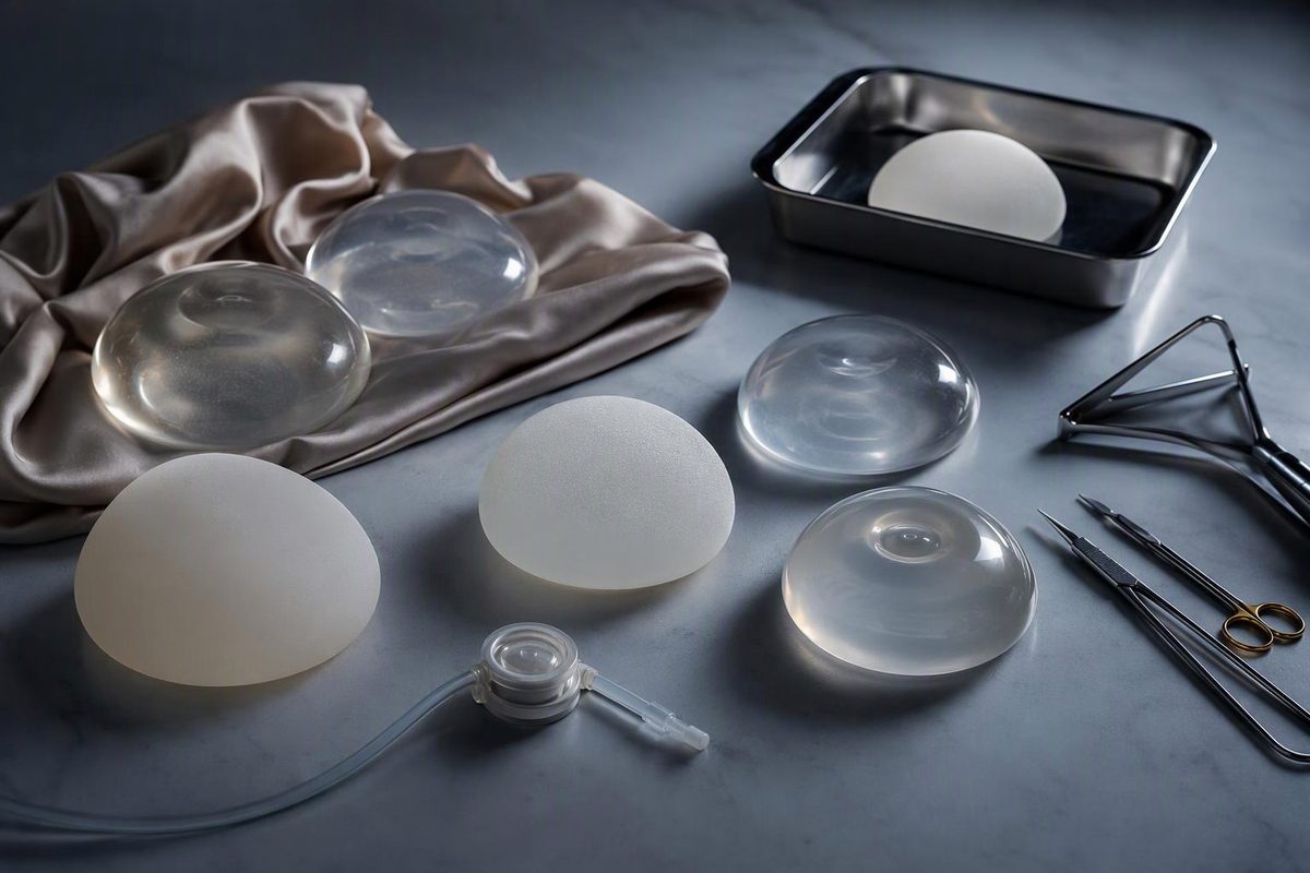
In dual-plane breast augmentation, the upper part of the implant sits behind the pectoralis major chest muscle while the lower part sits behind breast tissue. The aim is to combine more implant coverage at the top with less muscle constraint at the bottom.
Despite the name, the implant is not split between two sealed pockets. It occupies one continuous pocket: beneath muscle in the upper breast and beneath breast tissue lower down. Surgical commentary also notes that conventional “subpectoral” placement is usually only partially under the pectoralis, so surgeons do not always use dual plane and under the muscle consistently (Plastic and Reconstructive Surgery Global Open).
What Types I, II and III mean
The dual-plane type describes how far the surgeon separates the breast tissue from the front of the pectoralis muscle—not a standardized percentage of the implant covered by muscle.
| Technique | Tissue release in front of the muscle |
|---|---|
| Type I | No additional separation above the lower muscle edge |
| Type II | Separation extends to approximately the lower edge of the areola |
| Type III | Separation extends to approximately the upper edge of the areola |
In all three, the pectoralis origin is released along the inframammary fold—the crease under the breast—while upper muscle coverage is retained. Types II and III add progressively more separation between breast tissue and muscle. Type I has no added prepectoral dissection, which is why some authors question whether it is meaningfully different from conventional partial subpectoral placement (Plastic and Reconstructive Surgery Global Open).
Operative details and terminology vary. “Type II” on a clinic website is therefore less informative than a diagram showing the surgeon’s planned pocket, muscle release and breast-fold position.
Why a surgeon may recommend it
Dual-plane placement may be considered when the upper breast has limited natural tissue to conceal an implant. Muscle coverage can make the upper implant edge less visible and may reduce wrinkling compared with placement entirely in front of the muscle.
The lower release is intended to let breast tissue settle over the lower part of the implant. Surgeons may consider this approach for selected patients who have:
- thin upper-pole tissue that provides limited implant coverage;
- mild looseness or glandular sagging; or
- a constricted lower breast that needs expansion.
These are not automatic indications. Implant width and projection, tissue thickness, nipple position, skin stretch and the strength of the existing breast fold all affect the plan. The technique’s proposed effect on breast shape is also not settled: the cited surgical commentary questions whether the additional tissue separation produces a meaningful advantage over a well-executed partial subpectoral pocket.
Dual plane does not replace a breast lift
An implant adds volume; it does not reliably move a substantially low nipple or remove excess skin. A more extensive dual-plane release may change how mildly loose tissue sits over an implant, but it cannot be assumed to correct meaningful sagging.
Breast tissue can instead descend over an implant that remains relatively fixed, producing what surgeons call a waterfall or snoopy appearance. Published commentary on dual-plane surgery states that patients with glandular sagging may still need augmentation with mastopexy, or a breast lift, rather than an implant alone (Plastic and Reconstructive Surgery Global Open). A separate review defines the waterfall effect as breast tissue sliding over a fixed or encapsulated implant and describes mastopexy, sometimes with implant exchange, as a common corrective option when a patient wants treatment (Gland Surgery).
Ask where your nipple sits relative to the inframammary fold and what the surgeon predicts with an implant alone. If breast position—not only lost volume—is the concern, compare dual plane with the options in breast lift versus breast augmentation.
The main tradeoff: muscle coverage can mean movement
Because the upper implant remains under the pectoralis, contracting the chest can move or distort the breast. This is called breast animation deformity. It may appear only during forceful chest contraction, or it may be noticeable during ordinary activity.
Dual-plane placement does not eliminate this risk. A systematic review defined animation as a change or distortion in breast shape when the pectoralis contracts. It found only four eligible studies and concluded that animation appeared to increase with greater muscle involvement (Archives of Plastic Surgery). The studies used different operations, grading methods and patient groups, so their reported percentages should not be treated as a personal prediction.
Animation deserves particular discussion if your exercise, sport or work repeatedly engages the chest muscles. Ask to see comparable results both at rest and with the pectoralis flexed.
Other pocket-related tradeoffs include:
- Implant displacement: A 2019 meta-analysis found more displacement and animation with subpectoral than prepectoral augmentation. It found lower rates of capsular contracture and hematoma in the subpectoral group, with no significant difference in several other complications (Aesthetic Plastic Surgery). These findings compare broad pocket categories, not every modern dual-plane variation.
- Double bubble: The original breast crease can remain visible as a second line across the lower breast, particularly when a tight fold is lowered.
- Bottoming out: The implant can descend too far if the lower tissues or reconstructed fold do not hold it. In one retrospective dual-plane series, the authors linked both double bubble and bottoming out to fold anatomy and pocket dissection, showing why the technique’s label alone cannot predict the result (Aesthetic Plastic Surgery).
- Muscle-related recovery: Because the operation involves lifting and releasing part of the pectoralis, ask how that affects the surgeon’s restrictions for lifting, driving and chest exercise. Follow the operating surgeon’s instructions rather than a standard online timeline.
For a direct comparison with placement entirely in front of the muscle, see breast implants under vs over muscle.
Questions to take to the consultation
- What exactly do you mean by dual plane in my operation? Ask the surgeon to mark the planned muscle release and new breast fold on a diagram.
- Why does this pocket fit my tissue thickness and breast shape better than an over-muscle pocket?
- Would a lift address my loose skin or nipple position more predictably than an implant alone? Ask what each option can and cannot change.
- How might the result move when I flex my chest? Discuss your exercise and occupation, and request flexed as well as relaxed before-and-after views from comparable patients.
- How are implant dimensions being matched to my tissues? A larger implant is not a dependable substitute for a lift and adds weight to the lower breast.
- Will you lower my existing breast crease? If so, ask how the plan addresses double bubble and bottoming out.
- Which costs are excluded? The quote should identify anesthesia, facility fees, implants, postoperative care and the practice’s financial policy for revision. Breast implant cost factors explains why itemized totals are more useful than a surgeon-fee headline.
Pocket choice addresses only one part of implant surgery. The FDA advises that breast implants are not lifetime devices and that complications can lead to additional operations. Current required labeling includes a boxed warning, patient decision checklist and device-specific information that should be reviewed before surgery (FDA). Ask for the exact implant’s patient labeling, and keep the device card after the operation.